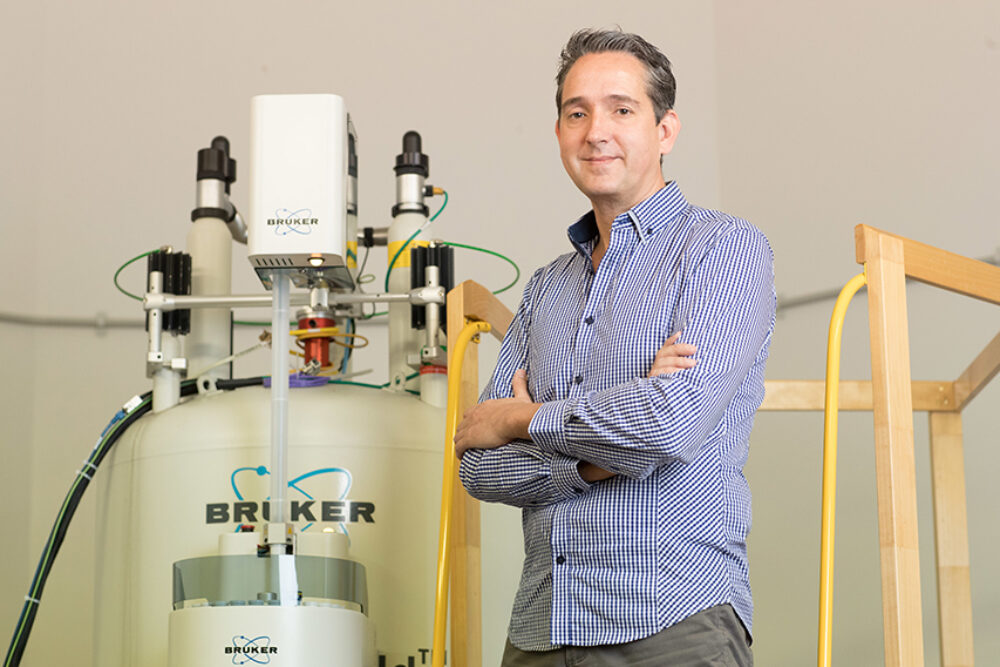
The Institute for Glycomics houses a wide range of cutting-edge facilities which aid our research scientists in their ongoing quest to take scientific discoveries from a laboratory setting and assess their potential to be translated into patient outcomes.
One of those facilities is our Nuclear Magnetic Resonance (NMR) Spectroscopy Facility. Our expert in this area, Associate Professor Thomas Haselhorst, explores NMR spectroscopy in detail; what it is, how it works, and why it’s important in our fight against some of the world’s most devastating diseases.
What is Nuclear Magnetic Resonance (NMR) Spectroscopy?
NMR is a prominent spectroscopy method for analysing compounds because it exploits the magnetic properties of certain atomic nuclei to determine the chemical and physical properties of these atoms or the molecules containing them.
It can provide extensive information about the structure, dynamics, and chemical environment of atoms. Additionally, NMR spectroscopy is ideally suited to distinguish different functional groups or identical functional groups in different molecular environments.
How does NMR Spectroscopy work?
In a typical NMR spectroscopy experiment, a thin glass tube containing a chemical compound, usually a liquid, is placed into a strong magnetic field.
All nuclei in the chemical compounds are in their ‘happy stage’ and align themselves either with or against the outside magnetic field. If we now ‘bombard’ these nuclei that are sitting in their ‘happy stage’ with a high frequency power pulse, they begin to ‘tumble’. During their ‘tumbling state’, the nuclei start to engage in all sorts of activity, e.g. they couple with each other, exchange magnetisation or perform a ‘handshake’ with neighbouring nuclei that are close in space, before they finally relax back into their ‘happy stage’.
All of this activity is recorded, and we obtain an NMR spectrum that contains important information about the chemical composition of this compound, purity, dynamic, transition states and even a three-dimensional structure of the molecule.

400 MHZ NMR SPECTROMETER 
600 MHZ NMR SPECTROMETER
How is it used and why is it important to biomedical research?
NMR spectroscopy is widely used in industry and academia and is the method of choice to identify novel compounds isolated from natural products or to validate synthetic strategy of a new chemical or drug.
NMR spectroscopy is also great for checking the purity of a new compound because, as I say to my students, “NMR never lies”. Every single molecule that is present in the glass tube will show up in an NMR spectrum. That means that, not only the compound you are interested in will show up in the NMR spectrum, but also impurities or by-products you don’t want in your sample.
However, NMR spectroscopy can do more than just compound validation. NMR is also a powerful technique to determine the three-dimensional structure of molecules such as proteins, glycans and DNA molecules.
How is NMR spectroscopy used in glycomics research?
Glycans are a particularly interesting class of molecules that can be investigated with NMR spectroscopy because glycans are inherently flexible molecules when they are dissolved in water and therefore difficult to study with other structural biology methods. NMR spectroscopy has the ability to monitor this flexibility and describe the structure and dynamic of glycans at an atomic level.
For example, we use NMR spectroscopy regularly to analyse the interaction of glycans with their target proteins, or cells, or whole intact viruses, bacteria or parasites. This is important to know, because the first thing that pathogens such as viruses, bacteria and parasites ‘see’ and interact with, are the glycans expressed on the host cells. Or, it is also possible, that the dense layer of glycans on their outer surface of many pathogens are used to ‘touch down’ and bind to the host cells to allow themselves entry into the body and thus causing disease. SARS-CoV-2 is an excellent example of viruses where glycans play a pivotal role in the cell recognition process.
Using NMR spectroscopy, we can exactly describe these important interactions at an atomic level. And if we know how pathogens such as viruses, bacteria or parasites enter the cell, we can design molecules that might inhibit this interaction and potentially prevent the disease.
Here at the Institute for Glycomics, we use NMR spectroscopy to also pinpoint the exact binding location of a glycan or drug when engaged with a target protein. For this experiment to work, the protein needs to be enriched with NMR active nuclei that we then monitor in the NMR spectrum.
The similarities between NMR and MRI (Magnetic Resonance Imaging)
NMR has proven to be of critical importance in the fields of medicine and chemistry, with new applications being developed daily.
NMR spectroscopy and MRI are very similar and rely on the same physical principle: Magnetic Resonance.
As described above, NMR spectroscopy works by placing a thin glass tube containing a chemical compound into a strong magnetic field, followed by the application of high frequency power pulses which excite the nuclei, causing them to ‘tumble’.
Most medical applications use Nuclear Magnetic Resonance Imaging, better known simply as Magnetic Resonance Imaging (MRI). MRI is an essential diagnostic tool to study the function and structure of the human body. It provides detailed images of any part of the body, especially soft tissue, in all possible planes and has been used in the areas of cardiovascular, neurological, musculoskeletal and oncological imaging.
In an MRI ‘experiment’, the human body takes over the role of the chemical compound in a glass tube. The patient will be horizontally inserted into the MRI scanner that also has an outside magnetic field. Because different places in the body contain different amounts of water, MRI detects the electromagnetic fields of the atoms in water molecules and uses this to determine differences in the density and shape of tissues throughout the body. This information can then be calculated into a 2D image that shows the details of your brain or body.
A very attractive MR application combining MRI and spectroscopy is the in-vivo MR spectroscopy, where chemicals and molecules in a particular area of the human body can be investigated, e.g. sugar molecules in brain or breast cancer. Chemical imbalances in the brain for patients with cancer or pain have been described.

How is NMR spectroscopy useful in cancer and infectious diseases research?
NMR is a very versatile technique in cancer research. The most important application of NMR in cancer research is the analysis of metabolites. Certain mutations in cancer cells can trigger a change in the overall metabolism of the body. If a certain compound or chemical is elevated, it might be an indication for the presence of cancer and NMR is ideal to identify the exact metabolite.
Other applications of NMR in cancer research are MRI and in-vivo MRS techniques as described above. NMR spectroscopy is also most essential in the development of novel anticancer drugs. Most anticancer drugs currently on the market would have undergone a rigorous NMR spectroscopic analysis to ensure the integrity of the compound.
The same is true for infectious diseases research and the majority of novel antiviral drugs would have been analysed by NMR spectroscopy. Interaction studies of host cell glycans with virus particles have been studied in the Institute by NMR spectroscopy, providing essential information on how the pathogen engages with the cell.
Our cutting-edge NMR facilities and professional expertise are available to external research groups and industry. To find out more, visit Our Facilities.
ABOUT THE AUTHOR

Associate Professor Thomas Haselhorst is a former Alexander von Humboldt Fellow and ARC Future Fellow and has international standing in structural glycoscience and NMR spectroscopy.
In particular, he has pioneered NMR spectroscopy techniques to elucidate interactions between glycan-based cell receptors and viruses, virus-like particles and intact cells.
Associate Professor Haselhorst has worked in the field of Rotavirus and Influenza virus research and recently in collaboration with Professor Michael Jennings and Dr Chris Day in SARS-CoV-2 drug discovery and structure-guided vaccine design.
Associate Professor Haselhorst’S research also includes the development of intelligent nanomaterials as targeted drug delivery systems for Non-Hodgkin’s Lymphoma and drug discovery of novel HIV and antifungal therapies.


